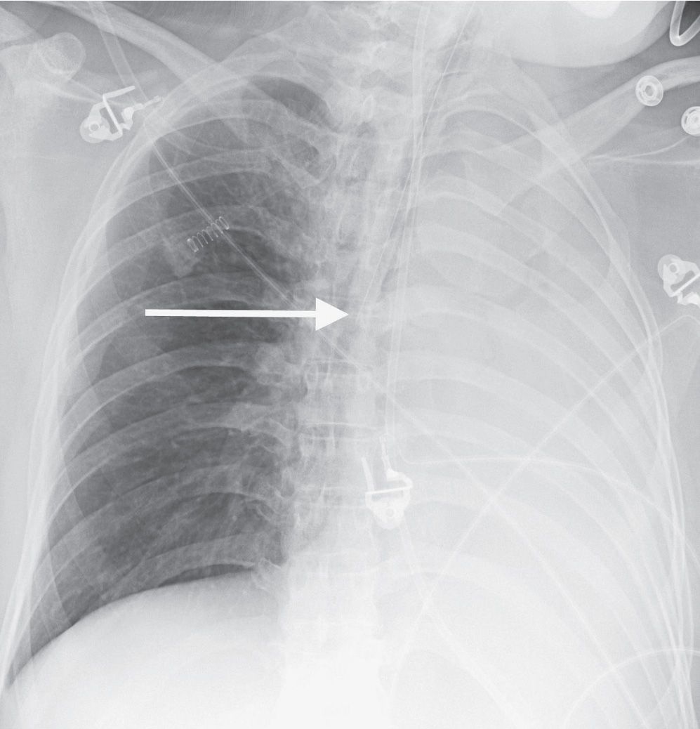
Chest X Ray Shows Atelectasis . how is atelectasis diagnosed? atelectasis describes the loss of lung volume due to the collapse of lung tissue. if imaging is warranted, chest radiography, chest computed tomography, or thoracic ultrasonography are useful when diagnosing. frontal and lateral views of the chest show an area of increased density (solid white arrow), which is silhouetting.
from ar.inspiredpencil.com
if imaging is warranted, chest radiography, chest computed tomography, or thoracic ultrasonography are useful when diagnosing. frontal and lateral views of the chest show an area of increased density (solid white arrow), which is silhouetting. atelectasis describes the loss of lung volume due to the collapse of lung tissue. how is atelectasis diagnosed?
Atelectasis Chest X Ray
Chest X Ray Shows Atelectasis if imaging is warranted, chest radiography, chest computed tomography, or thoracic ultrasonography are useful when diagnosing. frontal and lateral views of the chest show an area of increased density (solid white arrow), which is silhouetting. if imaging is warranted, chest radiography, chest computed tomography, or thoracic ultrasonography are useful when diagnosing. atelectasis describes the loss of lung volume due to the collapse of lung tissue. how is atelectasis diagnosed?
From www.animalia-life.club
Atelectasis Chest X Ray Chest X Ray Shows Atelectasis if imaging is warranted, chest radiography, chest computed tomography, or thoracic ultrasonography are useful when diagnosing. frontal and lateral views of the chest show an area of increased density (solid white arrow), which is silhouetting. atelectasis describes the loss of lung volume due to the collapse of lung tissue. how is atelectasis diagnosed? Chest X Ray Shows Atelectasis.
From www.shutterstock.com
X Ray Chest Atelectasis Stock Photo 1443126737 Shutterstock Chest X Ray Shows Atelectasis how is atelectasis diagnosed? frontal and lateral views of the chest show an area of increased density (solid white arrow), which is silhouetting. atelectasis describes the loss of lung volume due to the collapse of lung tissue. if imaging is warranted, chest radiography, chest computed tomography, or thoracic ultrasonography are useful when diagnosing. Chest X Ray Shows Atelectasis.
From www.shutterstock.com
Xray Chest Atelectasis Stock Photo (Edit Now) 1269376837 Chest X Ray Shows Atelectasis if imaging is warranted, chest radiography, chest computed tomography, or thoracic ultrasonography are useful when diagnosing. how is atelectasis diagnosed? atelectasis describes the loss of lung volume due to the collapse of lung tissue. frontal and lateral views of the chest show an area of increased density (solid white arrow), which is silhouetting. Chest X Ray Shows Atelectasis.
From www.researchgate.net
Chest Xray showing right pleural effusion with adjacent atelectasis Chest X Ray Shows Atelectasis atelectasis describes the loss of lung volume due to the collapse of lung tissue. if imaging is warranted, chest radiography, chest computed tomography, or thoracic ultrasonography are useful when diagnosing. how is atelectasis diagnosed? frontal and lateral views of the chest show an area of increased density (solid white arrow), which is silhouetting. Chest X Ray Shows Atelectasis.
From ar.inspiredpencil.com
Chest X Ray Atelectasis Chest X Ray Shows Atelectasis how is atelectasis diagnosed? atelectasis describes the loss of lung volume due to the collapse of lung tissue. if imaging is warranted, chest radiography, chest computed tomography, or thoracic ultrasonography are useful when diagnosing. frontal and lateral views of the chest show an area of increased density (solid white arrow), which is silhouetting. Chest X Ray Shows Atelectasis.
From ar.inspiredpencil.com
Chest X Ray Atelectasis Chest X Ray Shows Atelectasis if imaging is warranted, chest radiography, chest computed tomography, or thoracic ultrasonography are useful when diagnosing. how is atelectasis diagnosed? atelectasis describes the loss of lung volume due to the collapse of lung tissue. frontal and lateral views of the chest show an area of increased density (solid white arrow), which is silhouetting. Chest X Ray Shows Atelectasis.
From ar.inspiredpencil.com
Atelectasis Chest X Ray Chest X Ray Shows Atelectasis if imaging is warranted, chest radiography, chest computed tomography, or thoracic ultrasonography are useful when diagnosing. frontal and lateral views of the chest show an area of increased density (solid white arrow), which is silhouetting. atelectasis describes the loss of lung volume due to the collapse of lung tissue. how is atelectasis diagnosed? Chest X Ray Shows Atelectasis.
From www.researchgate.net
Chest Xray showed subsegmental bibasilar atelectasis Download Chest X Ray Shows Atelectasis how is atelectasis diagnosed? frontal and lateral views of the chest show an area of increased density (solid white arrow), which is silhouetting. atelectasis describes the loss of lung volume due to the collapse of lung tissue. if imaging is warranted, chest radiography, chest computed tomography, or thoracic ultrasonography are useful when diagnosing. Chest X Ray Shows Atelectasis.
From www.animalia-life.club
Atelectasis Chest X Ray Chest X Ray Shows Atelectasis atelectasis describes the loss of lung volume due to the collapse of lung tissue. how is atelectasis diagnosed? frontal and lateral views of the chest show an area of increased density (solid white arrow), which is silhouetting. if imaging is warranted, chest radiography, chest computed tomography, or thoracic ultrasonography are useful when diagnosing. Chest X Ray Shows Atelectasis.
From mungfali.com
Atelectasis Vs Pneumonia Chest X Ray Chest X Ray Shows Atelectasis frontal and lateral views of the chest show an area of increased density (solid white arrow), which is silhouetting. atelectasis describes the loss of lung volume due to the collapse of lung tissue. how is atelectasis diagnosed? if imaging is warranted, chest radiography, chest computed tomography, or thoracic ultrasonography are useful when diagnosing. Chest X Ray Shows Atelectasis.
From www.researchgate.net
Anteroposterior view of chest Xray shows basal atelectasis and Chest X Ray Shows Atelectasis how is atelectasis diagnosed? if imaging is warranted, chest radiography, chest computed tomography, or thoracic ultrasonography are useful when diagnosing. atelectasis describes the loss of lung volume due to the collapse of lung tissue. frontal and lateral views of the chest show an area of increased density (solid white arrow), which is silhouetting. Chest X Ray Shows Atelectasis.
From www.researchgate.net
Chest Xray showed atelectasis of the lower part of the left lung with Chest X Ray Shows Atelectasis atelectasis describes the loss of lung volume due to the collapse of lung tissue. frontal and lateral views of the chest show an area of increased density (solid white arrow), which is silhouetting. how is atelectasis diagnosed? if imaging is warranted, chest radiography, chest computed tomography, or thoracic ultrasonography are useful when diagnosing. Chest X Ray Shows Atelectasis.
From www.animalia-life.club
Atelectasis Chest X Ray Chest X Ray Shows Atelectasis atelectasis describes the loss of lung volume due to the collapse of lung tissue. how is atelectasis diagnosed? frontal and lateral views of the chest show an area of increased density (solid white arrow), which is silhouetting. if imaging is warranted, chest radiography, chest computed tomography, or thoracic ultrasonography are useful when diagnosing. Chest X Ray Shows Atelectasis.
From www.researchgate.net
Chest xray taken on admission showing left basilar atelectasis (red Chest X Ray Shows Atelectasis atelectasis describes the loss of lung volume due to the collapse of lung tissue. how is atelectasis diagnosed? frontal and lateral views of the chest show an area of increased density (solid white arrow), which is silhouetting. if imaging is warranted, chest radiography, chest computed tomography, or thoracic ultrasonography are useful when diagnosing. Chest X Ray Shows Atelectasis.
From www.animalia-life.club
Atelectasis Chest X Ray Chest X Ray Shows Atelectasis atelectasis describes the loss of lung volume due to the collapse of lung tissue. how is atelectasis diagnosed? if imaging is warranted, chest radiography, chest computed tomography, or thoracic ultrasonography are useful when diagnosing. frontal and lateral views of the chest show an area of increased density (solid white arrow), which is silhouetting. Chest X Ray Shows Atelectasis.
From www.shutterstock.com
Chest Xray Show Right Lung Atelectasis库存照片641043421 Shutterstock Chest X Ray Shows Atelectasis how is atelectasis diagnosed? if imaging is warranted, chest radiography, chest computed tomography, or thoracic ultrasonography are useful when diagnosing. frontal and lateral views of the chest show an area of increased density (solid white arrow), which is silhouetting. atelectasis describes the loss of lung volume due to the collapse of lung tissue. Chest X Ray Shows Atelectasis.
From www.researchgate.net
Chest CT shows hyperinflation of the RML, with pulmonary atelectasis Chest X Ray Shows Atelectasis atelectasis describes the loss of lung volume due to the collapse of lung tissue. frontal and lateral views of the chest show an area of increased density (solid white arrow), which is silhouetting. how is atelectasis diagnosed? if imaging is warranted, chest radiography, chest computed tomography, or thoracic ultrasonography are useful when diagnosing. Chest X Ray Shows Atelectasis.
From ar.inspiredpencil.com
Atelectasis Chest X Ray Chest X Ray Shows Atelectasis frontal and lateral views of the chest show an area of increased density (solid white arrow), which is silhouetting. if imaging is warranted, chest radiography, chest computed tomography, or thoracic ultrasonography are useful when diagnosing. atelectasis describes the loss of lung volume due to the collapse of lung tissue. how is atelectasis diagnosed? Chest X Ray Shows Atelectasis.
